Doctors and Dissection
A selection of prints relating to the exhibition Doctors, Dissection & Resurrection Men currently at the Museum of London.
One of the most famous representations of dissection in art is the final plate from William Hogarth's Four Stages of Cruelty, in which Hogarth's antagonist Tom Nero gets his comeuppance. The following illustration is from a set of copies of Hogarth's print, published with permission shortly after Hogarth's death:
 [The four stages of cruelty]
Design'd by W.m Hogarth.
[London: Robert Sayer, 1767.]
Set of four engravings, with large margins. Each 360 x 260mm, 14¼ x 10¼".
The progress of Tom Nero from his childhood torturing of a dog to murder, the gallows and the dissection table. After Hogarth's death in 1764 his widow Jane petitioned Parliament for extra copyright and was granted an additional twenty years of exclusivity. However she gave the London publisher Robert Sayer permission to publish 'Les Satyres de Guillaume Hogarth Oeuvre Moral et Comique', one of the earliest authorized collections of Hogarth's work.
[Ref: 26607] £280.00
Hogarth's dissection is said to have been inspired by the following print after Dutch-English artist Egbert van Heemskerck:
 Behold how in the colledge hall, the surgeons and the doctors all, Are met in consultation wise, a carcase to anatomize: the master there displays his art, sagely discants on every part, and that with ears & eyes and nose, we hear, and see, and smell, he shows.
[Drawn by Egbert van Heemskerck.]
[n.d,. engraved c.1730, but printed c.1800.)
Copper engraving. 290 x 250mm, 11½ x 9¾". Large margins. Paper brittle, backed on thin tissue.
[Ref: 19723] £280.00
An earlier continental dissection can be seen here:

[Headpiece?: animal dissection a the Academie des sciences, Paris.]
Le Clerc in. et fecit.
[Paris: Imprimerie royale?, n.d., c.1670s.]
Etching, illustration to a book, sheet/etched frame 100 x 245mm, 4 x 9¾". A fragment, with French letterpress to verso, rare. No margin outside border; staining upper right.
A wolf or fox's(?) internal organs are examined around a table by scientists; some consult charts and open books. Other learned figures conversing in groups behind, human and animal skeletons and skulls against the far wall.
[Ref: 27669] £260.00
In the 18th century, lavish books of anatomical illustrations were published to assist lecturers and students. This print is from the famous Cours complet d'anatomie by Gautier d'Agoty, another plate from which is in the Museum of London's exhibition:
 [Cours Complet d'Anatomie.]

This volume covered all stages of preganancy, and was authored by William Hunter (1718-83), the physician, anatomist, and man-midwife.
William Hunter was the elder brother of John Hunter (1728-93), also an anatomist and remembered as a founder of 'scientific surgery'. John Hunter's collection was purchased by the government after his death, and can be seen to this day at the Hunterian Museum in London, which celebrates its bicentenary this year. Hunter was painted by his Leicester Square neighbour Sir Joshua Reynolds in 1786, surrounded by his publications and specimens from his collection, a portrait which 'became his public image, thanks to the hundred or more impressions of William Sharp's even more expressive print' (DNB), as seen here:

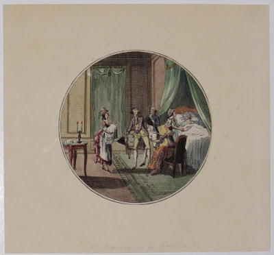
The nineteenth century presentation of anatomical specimens can also be seen in this depiction of the Museo di Anatomia Umana in Rome:

Around the turn of the nineteenth century, the business of instructing anatomy was flourishing- this print, advertising the anatomy lectures of the Scottish surgeons John Bell (1763-1820) and his brother Charles (1774-1842), combines the developments in scientific method with an image (designed by John Bell himself!) indebted to the iconography of the Romantics:

A crucial moment for anatomy in the nineteenth century was the case of the 'Italian Boy', Carlo Ferrari (for more on which see Sarah Wise's excellent book, 'The Italian Boy'). Ferrari's death, for which the 'Resurrectionists' May, Bishop and Williams were tried, brought to the public's attention the ways in which bodies were procured for dissection, often though graverobbing or outright murder, often with the collusion of unscrupulous anatomy schools. As the Museum of London exhibition shows, a plethora of printed images were produced to represent and idealise the Italian Boy, including these:

In the wake of the outrage provoked by this case, the 1832 Anatomy Act was passed, to give freer licence to hospitals and anatomists to dissect unclaimed and donated bodies, increasing the supply of bodies available for dissection. This was in order to dissuade them from colluding with criminals to augment the meagre supply of bodies hitherto legally available (strictly speaking, only those of executed criminals).

One of the most famous representations of dissection in art is the final plate from William Hogarth's Four Stages of Cruelty, in which Hogarth's antagonist Tom Nero gets his comeuppance. The following illustration is from a set of copies of Hogarth's print, published with permission shortly after Hogarth's death:

Design'd by W.m Hogarth.
[London: Robert Sayer, 1767.]
Set of four engravings, with large margins. Each 360 x 260mm, 14¼ x 10¼".
The progress of Tom Nero from his childhood torturing of a dog to murder, the gallows and the dissection table. After Hogarth's death in 1764 his widow Jane petitioned Parliament for extra copyright and was granted an additional twenty years of exclusivity. However she gave the London publisher Robert Sayer permission to publish 'Les Satyres de Guillaume Hogarth Oeuvre Moral et Comique', one of the earliest authorized collections of Hogarth's work.
[Ref: 26607] £280.00
Hogarth's dissection is said to have been inspired by the following print after Dutch-English artist Egbert van Heemskerck:
[Drawn by Egbert van Heemskerck.]
[n.d,. engraved c.1730, but printed c.1800.)
Copper engraving. 290 x 250mm, 11½ x 9¾". Large margins. Paper brittle, backed on thin tissue.
[Ref: 19723] £280.00
An earlier continental dissection can be seen here:

[Headpiece?: animal dissection a the Academie des sciences, Paris.]
Le Clerc in. et fecit.
[Paris: Imprimerie royale?, n.d., c.1670s.]
Etching, illustration to a book, sheet/etched frame 100 x 245mm, 4 x 9¾". A fragment, with French letterpress to verso, rare. No margin outside border; staining upper right.
A wolf or fox's(?) internal organs are examined around a table by scientists; some consult charts and open books. Other learned figures conversing in groups behind, human and animal skeletons and skulls against the far wall.
[Ref: 27669] £260.00
In the 18th century, lavish books of anatomical illustrations were published to assist lecturers and students. This print is from the famous Cours complet d'anatomie by Gautier d'Agoty, another plate from which is in the Museum of London's exhibition:

Gautier D'agoty fils secon pinx.
[Paris, n.d, 1773.]
Coloured engraving, 390 x 540mm. Staining right hand corner, damage to top right.
Plan XV. Showing chest and pelvis anatomy. From the first series of large scale coloured engravings of anatomy.
[Ref: 2658] £820
[Paris, n.d, 1773.]
Coloured engraving, 390 x 540mm. Staining right hand corner, damage to top right.
Plan XV. Showing chest and pelvis anatomy. From the first series of large scale coloured engravings of anatomy.
[Ref: 2658] £820
This plate of the uterus and blood supply to the placenta, is from a drawing by Jan van Rymsdyck, and was published in the Anatomia uteri humani gravidi tabulis illustrata, (1774):

[Tab. XVII. Idem Uterus a fronte Primo sive extimo substantiae strato sublato vasorum ampliorum distributio, corumque versus Placentam (quae hic parti anteriori et fundo Uteri adhaerebat) progressus melius conspiciuntur.] [J.V. Rymsyke delin. Menil sculp.]
[Birmingham: John Baskerville, 1774.]
Copper engraving. Plate 437 x 299mm. 17¼ x 11¾". Linen-backed for lecture use with original ties to hang-up. Text removed, abraised. Large water-stain to right.
[Ref: 20910] £260
[Birmingham: John Baskerville, 1774.]
Copper engraving. Plate 437 x 299mm. 17¼ x 11¾". Linen-backed for lecture use with original ties to hang-up. Text removed, abraised. Large water-stain to right.
[Ref: 20910] £260
This volume covered all stages of preganancy, and was authored by William Hunter (1718-83), the physician, anatomist, and man-midwife.
William Hunter was the elder brother of John Hunter (1728-93), also an anatomist and remembered as a founder of 'scientific surgery'. John Hunter's collection was purchased by the government after his death, and can be seen to this day at the Hunterian Museum in London, which celebrates its bicentenary this year. Hunter was painted by his Leicester Square neighbour Sir Joshua Reynolds in 1786, surrounded by his publications and specimens from his collection, a portrait which 'became his public image, thanks to the hundred or more impressions of William Sharp's even more expressive print' (DNB), as seen here:
John Hunter.
Sir Joshua Reynolds pinx.t Will.m Sharp sculp.t
London, Published 1.st Jan.y 1788, by W.m Sharp, No.8, Charles Street, Midd.x Hospital: B.B. Evans, corner of the Old Jewry, Cheapside, & W. Skelton, No.23 Hay Market.
Copper engraving.
Hamilton: p.39. Wellcome: 1475-4.
[Ref: 26504] £480
Sir Joshua Reynolds pinx.t Will.m Sharp sculp.t
London, Published 1.st Jan.y 1788, by W.m Sharp, No.8, Charles Street, Midd.x Hospital: B.B. Evans, corner of the Old Jewry, Cheapside, & W. Skelton, No.23 Hay Market.
Copper engraving.
Hamilton: p.39. Wellcome: 1475-4.
[Ref: 26504] £480
The nineteenth century presentation of anatomical specimens can also be seen in this depiction of the Museo di Anatomia Umana in Rome:
Museo di Anatomia Umana nell'Archiginnasio Romano.
[P. Cacchiatelli. G. Cleter.]
[Roma, Tip. delle Belle Arti.] [n.d. c.1865.]
Sepia aquatint, with large margins. Plate 240 x 285mm. 9½ x 11¼". Crease through centre.
[P. Cacchiatelli. G. Cleter.]
[Roma, Tip. delle Belle Arti.] [n.d. c.1865.]
Sepia aquatint, with large margins. Plate 240 x 285mm. 9½ x 11¼". Crease through centre.
[Ref: 26849] £160.00
(£192.00 incl.VAT)
Around the turn of the nineteenth century, the business of instructing anatomy was flourishing- this print, advertising the anatomy lectures of the Scottish surgeons John Bell (1763-1820) and his brother Charles (1774-1842), combines the developments in scientific method with an image (designed by John Bell himself!) indebted to the iconography of the Romantics:

Lectures on Anatomy & Surgery. Iohn and Charles Bell. "Wherefore is there a price in the hand of a fool to get Wisdom seeing he hath no heart to it".
J. Bell invent. et delin. J. Neagle sculp.
[n.d. c.1800.]
Engraving, rare with large margins. Plate 178 x 210mm. 7 x 8¼". Tear into upper edge; soiling and foxing around extremeties.
[Ref: 26122] £160.00 (£192.00 incl.VAT)
J. Bell invent. et delin. J. Neagle sculp.
[n.d. c.1800.]
Engraving, rare with large margins. Plate 178 x 210mm. 7 x 8¼". Tear into upper edge; soiling and foxing around extremeties.
[Ref: 26122] £160.00 (£192.00 incl.VAT)
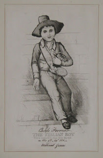
A crucial moment for anatomy in the nineteenth century was the case of the 'Italian Boy', Carlo Ferrari (for more on which see Sarah Wise's excellent book, 'The Italian Boy'). Ferrari's death, for which the 'Resurrectionists' May, Bishop and Williams were tried, brought to the public's attention the ways in which bodies were procured for dissection, often though graverobbing or outright murder, often with the collusion of unscrupulous anatomy schools. As the Museum of London exhibition shows, a plethora of printed images were produced to represent and idealise the Italian Boy, including these:

The Italian Boy Who is supposed to have been Burked. [In Ink:] Carlo Ferriar on the 4th Novr. 1831 in Bethnal Green.
[n.d. c.1831.]
A rare aquatint. 247 x 158mm.
[Ref: 18416] £140.00 (£168.00 incl.VAT)
[n.d. c.1831.]
A rare aquatint. 247 x 158mm.
[Ref: 18416] £140.00 (£168.00 incl.VAT)

The Italian Boy. Supposed to have been assassinated_whose body was sold for dissection at the Kings College. See Times Nov. 8th 1831
Painted, & Litho: by W. Franquinet. W. Day Lith.r to the King, 17, Gate St.
Pub.d by H. Lacy, 1 Wells Street._ Oxford Street
Lithograph, sheet 215 x 180mm (8½ x 7"). [Ref: 26928] £120.00 (£144.00 incl.VAT)
Painted, & Litho: by W. Franquinet. W. Day Lith.r to the King, 17, Gate St.
Pub.d by H. Lacy, 1 Wells Street._ Oxford Street
Lithograph, sheet 215 x 180mm (8½ x 7"). [Ref: 26928] £120.00 (£144.00 incl.VAT)
In the wake of the outrage provoked by this case, the 1832 Anatomy Act was passed, to give freer licence to hospitals and anatomists to dissect unclaimed and donated bodies, increasing the supply of bodies available for dissection. This was in order to dissuade them from colluding with criminals to augment the meagre supply of bodies hitherto legally available (strictly speaking, only those of executed criminals).
Macaronies Drawn After the Life. V.2. 22.
Pub. Accord to Act Dec.r 1. 1773 by MDarly 39 Strand.
Etching with small margins, printed on 18th century watermarked paper. Plate 178 x 247mm. 7 x 9¾".
BM Satires: 4645.
[Ref: 27853] £320
Pub. Accord to Act Dec.r 1. 1773 by MDarly 39 Strand.
Etching with small margins, printed on 18th century watermarked paper. Plate 178 x 247mm. 7 x 9¾".
BM Satires: 4645.
[Ref: 27853] £320